Precision Planning Starts With a Clearer Picture
How is a periodontist able to place a dental implant with precision inches from major nerves and sinus cavities without directly viewing them during surgery? The answer lies in imaging technology that goes far beyond a standard dental X-ray. A flat, two-dimensional X-ray can show the location of a tooth, but it cannot reveal the exact height, width, and density of the bone beneath the gums.
At Harmony Cosmetic Dentistry & Implants, we use 3D Cone Beam Computed Tomography (CBCT) to evaluate that hidden anatomy before any surgical instrument is used. This single scan gives Dr. Haider Alramli a complete map of the jaw, teeth, and sinuses, allowing him to plan dental implants, bone grafts, and other procedures with a level of precision that flat imaging cannot match.
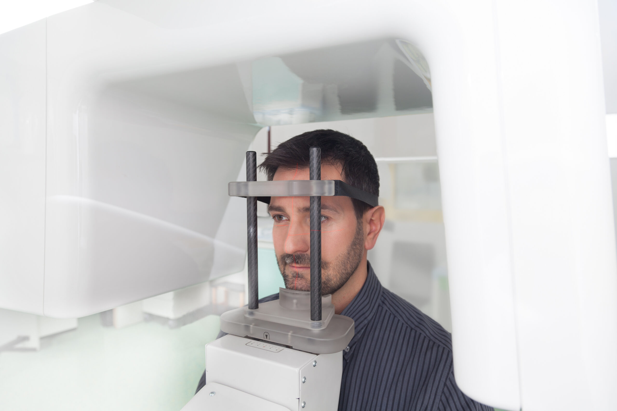
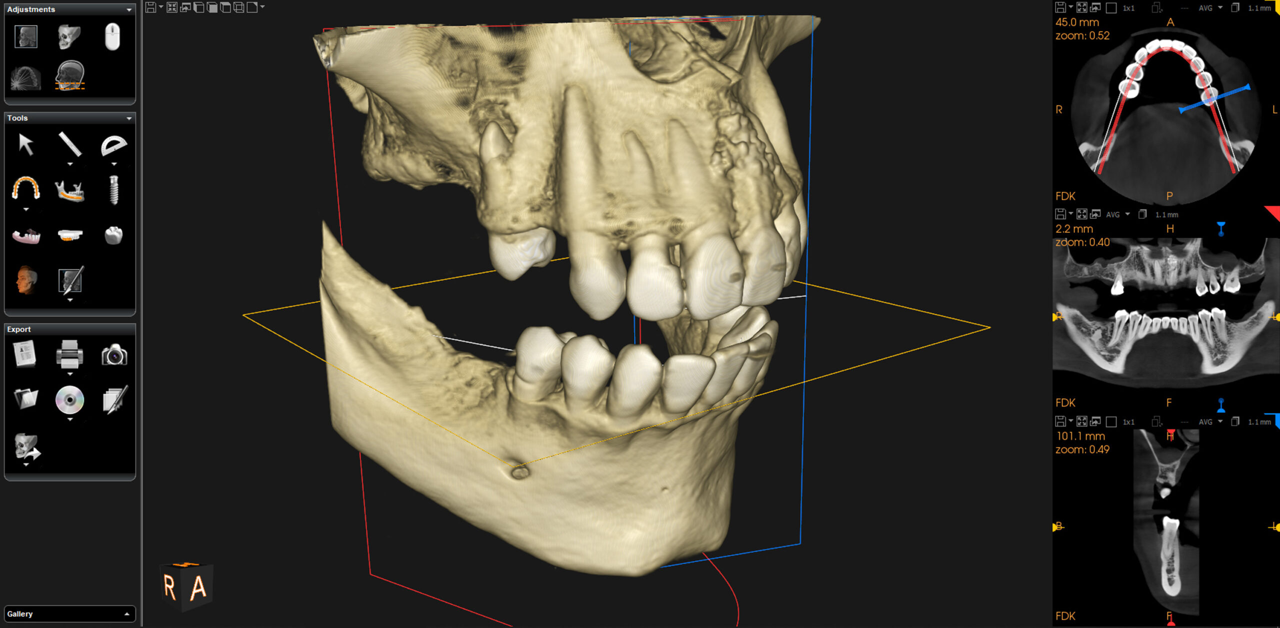
Cone Beam Computed Tomography is a specialized scanner that rotates around your head, capturing a series of images that are then reconstructed into a detailed 3D model of your teeth, jawbone, and sinuses. Unlike a traditional X-ray, which produces a single flat picture, CBCT captures volumetric data, meaning we can rotate, zoom, and slice through the image in any direction to examine a specific area from every angle.
Dental CBCT allows us to see anatomy that a conventional two-dimensional X-ray cannot reveal, giving Dr. Haider a far more complete picture for diagnosis and treatment planning. Rather than estimating based on a flat image, we can view your bone structure, tooth roots, and sinus cavities from any angle before a single decision is made about your care.
Before placing a dental implant, Dr. Haider uses your CBCT scan to identify the strongest zones of your jawbone. This allows him to plan the exact position and angle of each implant while avoiding important nerves and blood vessels, giving you a safer, more predictable surgical outcome.

Your scan reveals precisely how much bone remains and how close your sinus floor sits to the treatment area. This information helps Dr. Haider determine whether a sinus lift or bone graft is needed to build a stable foundation before implant placement.
CBCT can also map the internal anatomy of a tooth in three dimensions, supporting more accurate diagnosis for complex root canal cases. For full-arch restorations, the same scan helps identify the densest bone available to support multiple implants at precise angles.
If your treatment plan calls for a 3D scan, the process is simple and typically adds only a few minutes to your visit. Here is what each step looks like from start to finish.
You will be asked to remove any metal jewelry, glasses, or removable dental appliances that could interfere with the image. This step helps ensure the scan captures an accurate picture of your teeth and jaw before we begin.
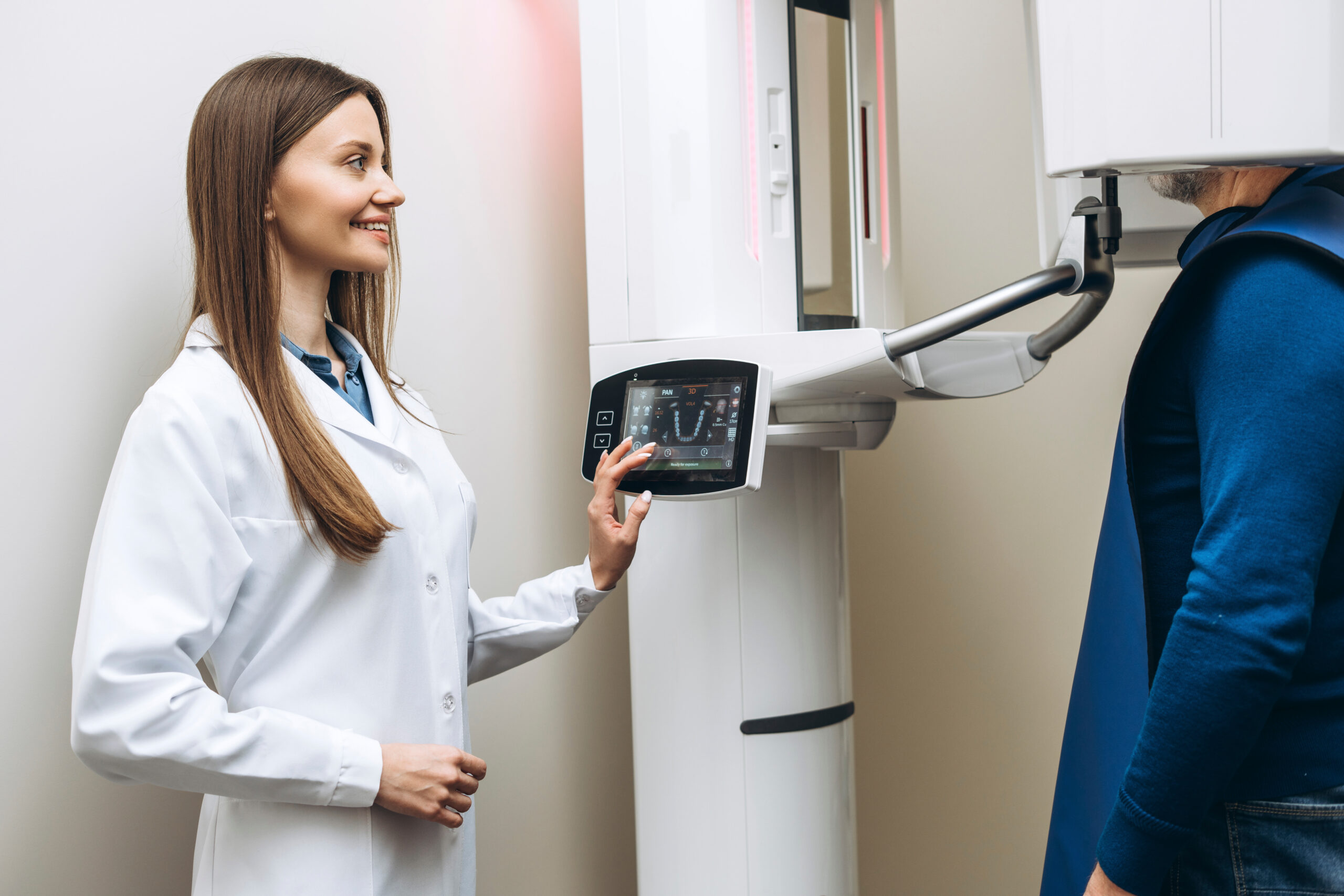
You will stand or sit comfortably near the scanner while it rotates around your head. There is no need to lie down inside an enclosed tube, and the open design helps most patients feel at ease throughout the process.
The scanner completes its full rotation in only a few seconds, capturing the volumetric data needed to build your 3D model. Because the images are recorded digitally, they appear on our screen almost instantly once the scan is complete.
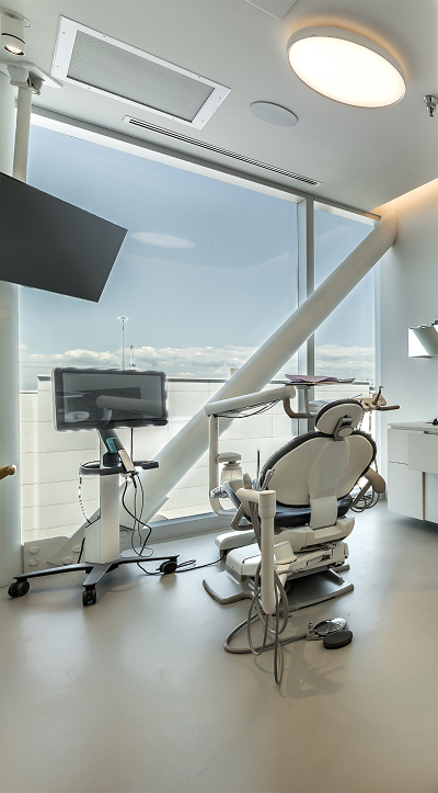
Dr. Haider reviews the images with you directly, pointing out the anatomy relevant to your procedure and what it means. This conversation helps you feel confident in your treatment plan before moving forward.
Whether you are exploring dental implants, need a bone graft evaluated, or simply want to understand what is happening beneath the surface of your smile, our Magnolia team is here to walk you through the process in clear, understandable terms. To learn more about how 3D imaging factors into your specific treatment needs, contact our office to schedule a consultation with our team.
Located in Magnolia, TX
Mon - Sat: 8AM to 5PM Sun: Closed